CARDIOVASCULAR ANATOMY
Blood flow through the heart: chambers, valves and circulation
Trace the path from the body to the lungs and back, with a labelled flow diagram, a heart cutaway and clinical connections.
Explore the heart and vascular atlas ↗One circuit, two pumps
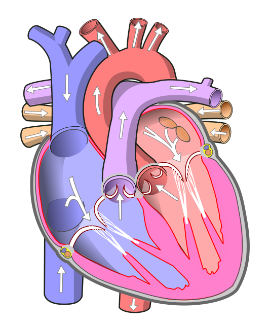
The right ventricle sends blood to the lungs for gas exchange. The left ventricle sends blood into the systemic circulation. Follow the arrows through both sides before returning to the starting point. [1]
The four valves in order
| Valve | Forward flow |
|---|---|
| Tricuspid | Right atrium → right ventricle |
| Pulmonary | Right ventricle → pulmonary trunk |
| Mitral | Left atrium → left ventricle |
| Aortic | Left ventricle → aorta |
Valves help prevent backward flow. Arteries carry blood away from the heart; veins carry blood toward it. Pulmonary arteries therefore carry oxygen-poor blood, while pulmonary veins carry oxygen-rich blood. [1]
.svg)
Clinical connections
Valve narrowing
Stenosis restricts forward flow through a valve. Identify the chamber just upstream of the narrowed opening. [2]
Valve leakage
Regurgitation allows reverse flow through a valve that does not close effectively. Add a backward arrow to the affected connection. [2]
Coronary circulation
The heart muscle needs its own blood supply. Coronary arteries branch from the aorta; blood within the chambers is not its arterial supply. [1]
Practise the route
Start with a red blood cell returning from a leg. Trace every chamber and valve until it reaches the aorta. Then repeat from a pulmonary vein. Use the diagram to check the sequence before exploring the animated flow routes.
Open CardioAtlas ↗References
- NHLBI: How blood flows through the heart — normal flow, valves and coronary supply.
- NHLBI: Heart valve diseases — stenosis and regurgitation.