Conceptual image-formation schematic · not to scale
A volume from measurements around the body
CT reconstructs cross-sectional information from X-ray measurements acquired at multiple angles. A slice places structures in a plane rather than superimposing the entire body thickness as one projection.
Stored value versus displayed shade
Hounsfield units describe attenuation on a calibrated CT scale. Window level selects the centre of a display range; width controls its span. Changing the window changes how stored values map to grayscale, not the original values.
FICTIONAL WORKED SCENARIO
Water is the same insert in two windows
The lab assigns the water insert 0 HU. At level 40 and width 400, it maps to approximately 102/255. At level −600 and width 1500, it maps to approximately 230/255. It looks lighter while its assigned value remains 0 HU.
The model uses the DICOM default LINEAR function. Rounding produces an 8-bit display.
Use the lab deliberately
- Choose soft tissue and locate the five inserts in the synthetic phantom.
- Narrow the width. Notice that a smaller interval uses the entire grayscale.
- Move level. Notice which inserts clip to black or white.
Clinical context
Window presets are display choices, not acquisition protocols. Contrast timing and reconstruction choices also matter. A fixed JPEG cannot restore original HU values, missing slices or information absent from an acquisition. CT uses ionizing radiation; contrast decisions need agent-specific assessment.
A real image to inspect
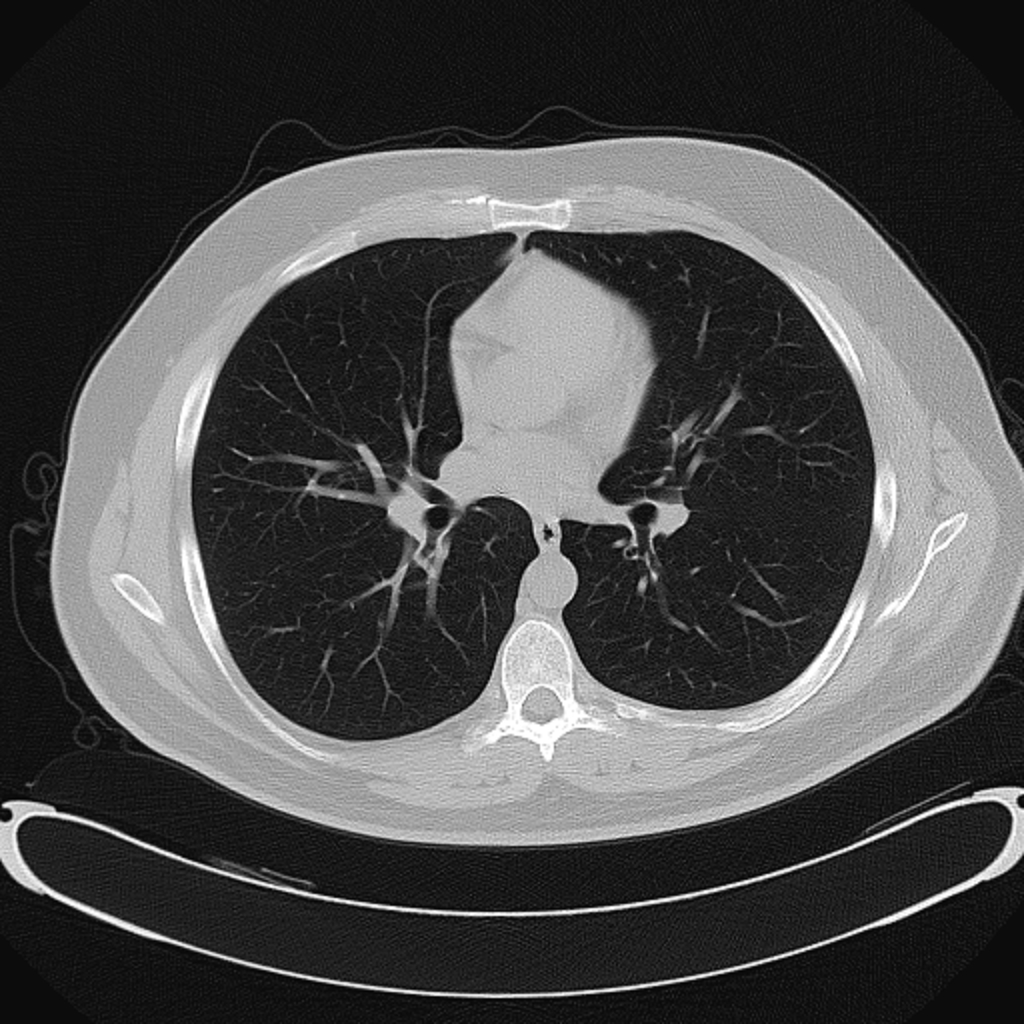
Chest CT in a lung window
An axial chest CT displayed in a lung window, showing branching pulmonary vessels against air-filled lungs. This is a fixed display image, not raw CT data; it cannot support true Hounsfield-unit windowing.
Ptrump16 · CC BY-SA 4.0 · Unchanged

Independent teaching image; not the patient from the fictional scenario.