Conceptual image-formation schematic · not to scale
Read a projection as a sum along a path
A radiograph combines contributions from everything the beam passes through. Two objects at different depths can occupy the same place in the resulting image. That makes an extra view useful: a changed angle can separate structures that overlapped.
A three-step reading habit
- Check the view. Identify projection and positioning before comparing apparent size.
- Describe the appearance. Use location, brightness and shape before naming a disease.
- Name the limitation. Ask what could be hidden by overlap or poorly depicted soft tissue.
FICTIONAL WORKED SCENARIO
The heart looks larger on a portable image
An earlier PA chest image and a later portable AP image show different apparent cardiac sizes. A change in silhouette alone cannot establish true enlargement. Projection geometry can contribute to magnification.
Make the geometry visible
In the lab, move the object away from a detector while keeping the source-to-detector distance fixed. Its shadow enlarges. This isolates one geometric effect; real radiographs also depend on positioning, inspiration and acquisition technique.
Clinical context
Radiographs commonly examine the chest and skeleton. They use ionizing radiation. A normal bone image does not exclude every soft-tissue injury; image selection and interpretation depend on the clinical question.
A real image to inspect
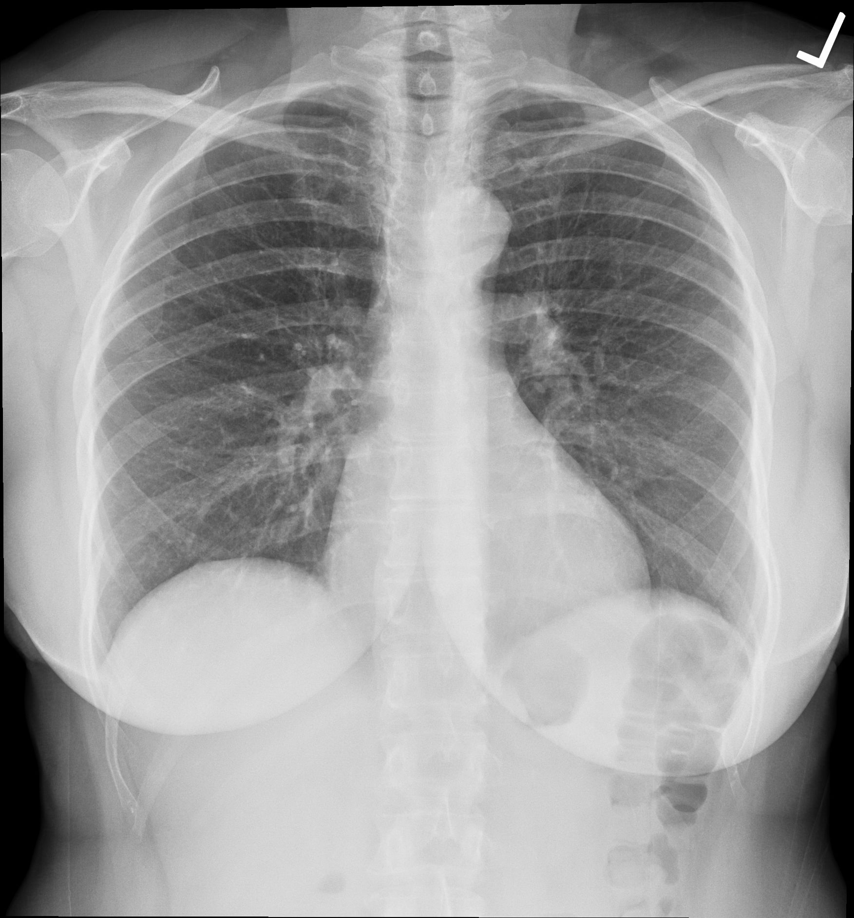
Chest radiograph
Frontal chest radiograph showing the lungs, cardiac silhouette and ribs. The source identifies this as a normal digitally acquired chest radiograph.
Ptrump16 · CC BY-SA 4.0 · Unchanged

Independent teaching image; not the patient from the fictional scenario.